Case Studies in Comprehensive Rhinoplasty
These case studies highlight real surgical outcomes achieved by Dr. Sam Most using patient-specific approaches that combine structural support and preservation techniques. Each case includes before-and-after photos, procedural details, and the rationale behind surgical decisions.
Case 1: Dorsal Hump Reduction with Tip Support
Patient Overview: This patient presented with a large dorsal hump and a prominent nasal profile. The surgical plan focused on traditional hump reduction using conventional resection techniques, combined with structural grafting and reshaping for long-term symmetry and support.
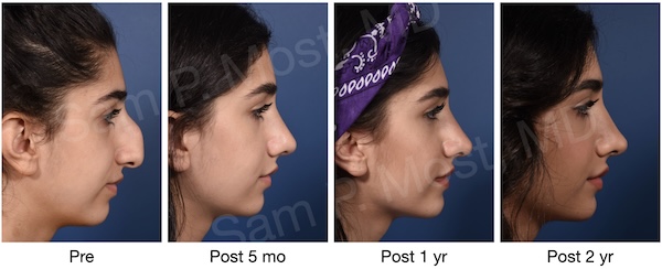
Fig. 17-1: Before and progressive after lateral images of a large dorsal hump reduction using conventional hump resection.
Surgical Technique: The procedure was performed via an external rhinoplasty approach. ULCs were separated from the septum, and the bony dorsum was addressed using standard and piezo rasping. Septoplasty preserved a 1.2 cm L-strut. Spreader grafts were placed (double-layered on the right) for midvault reconstruction. Cephalic trims preserved 8 mm of the LLCs, and a 4 mm lateral crural overlay was performed with strut grafts to prevent collapse. Tongue-in-groove technique set the medial crura and adjusted rotation. An alar-spanning suture was placed. SNAS fascia and crushed cartilage were added for dorsal contour symmetry.
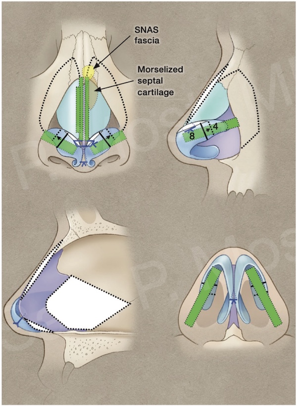
Fig. 17-3: Diagram of procedures performed including grafts and osteotomies.
Case 2: Dorsal Preservation Using MSSM
Patient Overview: This patient underwent dorsal preservation rhinoplasty using Dr. Most’s Modified Subdorsal Strip Method (MSSM), aiming to reduce dorsal projection while maintaining natural contour lines.
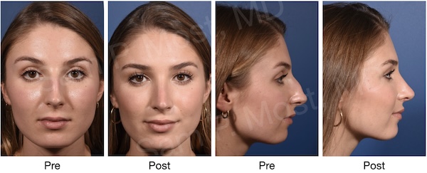
Fig. 18-1: Before and after (1 year) views of a patient who underwent dorsal preservation using MSSM. Notice the natural appearance.
Surgical Technique: A let-down dorsal preservation with full open approach was used. Piezo-assisted lateral ostectomies allowed dorsal mobilization. A subdorsal strip incision 6 mm below the dorsum preserved a cartilage segment for controlled lowering. ULCs were released laterally. The subdorsal strut was sutured to a preserved 1.3 cm caudal strut. An alar-spanning suture ensured dome symmetry.
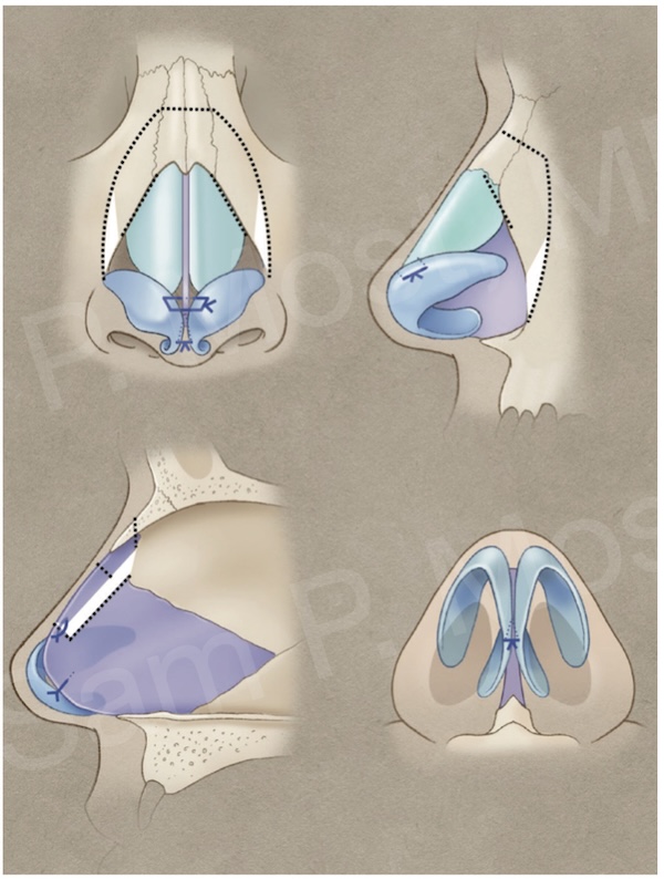
Fig. 18-3: Diagram of let-down dorsal preservation with MSSM technique.
Case 3: Saddle Nose Repair with Rib Grafting
Patient Overview: This patient had a severe saddle nose deformity due to collapse of the midvault and loss of upper lateral cartilage support. Surgery aimed to restore both structure and function.
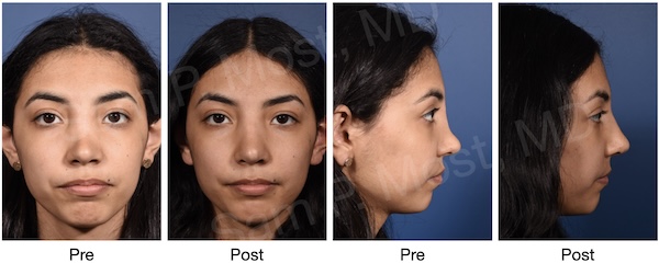
Fig. 19-1: Before and 14 month postoperative views after functional repair of a saddle nose deformity.
Surgical Technique: Via an external approach, absent ULCs and overly long nasal bones were addressed. A dorsal onlay spreader graft was placed and secured after bone release. ASR with rib cartilage was fixed with a miniplate to the premaxilla. Tongue-in-groove and alar-spanning sutures controlled tip position. A dorsal onlay diced rib/fibrin glue graft provided camouflage.
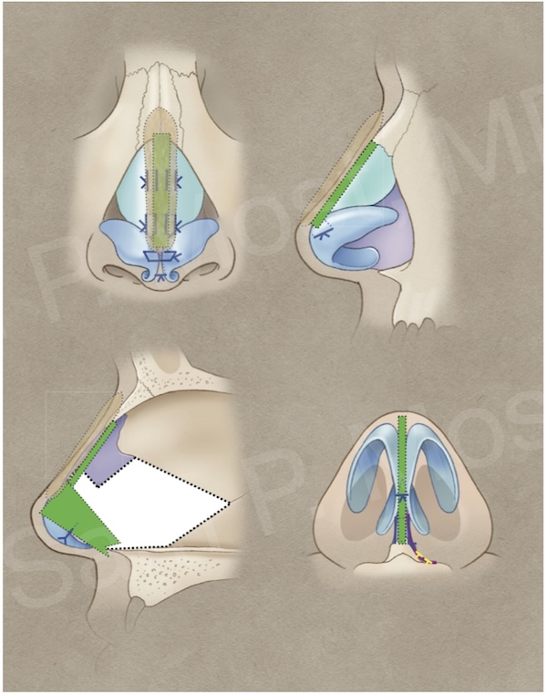
Fig. 19-3: Diagram of procedures including rib grafting, ASR, and diced cartilage onlay.
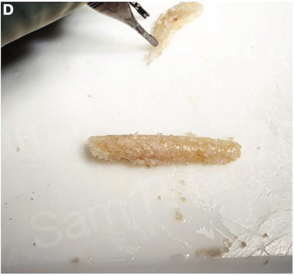
Fig. 19-4: Example of diced rib/fibrin glue graft (not from this case).
Case 4: Caudal Tetrapod Reconstruction for Short Nose
Patient Overview: A patient with a short nose and midvault asymmetry underwent caudal extension to increase nasal length and frontal symmetry.
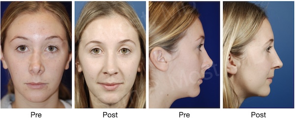
Fig. 20-1: Before and after (11 month) views showing improved frontal symmetry after caudal tetrapod reconstruction.
Surgical Technique: Septal cartilage was harvested for a right-sided SEG. A percutaneous right osteotomy and left-sided spreader graft were performed. Morselized cartilage enhanced dorsal symmetry. The tongue-in-groove method stabilized medial crura. Lateral crural remnants were replaced with strut grafts placed in precise, lower pockets.
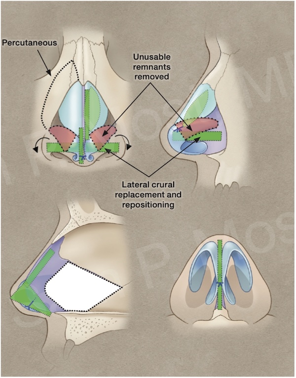
Fig. 20-2: Diagram of SEG, dorsal augmentation, and tip reconstruction.
Conclusion
These cases illustrate how functional and aesthetic goals can be harmonized using individualized strategies. Whether through dorsal preservation, rib graft reconstruction, or revision of prior surgery, Dr. Most applies consistent principles of structure, support, and preservation to achieve long-lasting and natural results.